


Breast cancer is still the most dangerous oncology for the female half of the planet. The most effective methods of early diagnosis are imaging tests such as mammography, ultrasound, MRI, etc. So what happens if the doctor does see an anomaly in the image? Firstly, there is no need to panic: most women who need additional follow-up examinations after a mammogram are healthy. Usually, when detecting neoplasms, the doctor prescribes additional ultrasound, and only 1-2% of patients will need a biopsy. So, what types of biopsy are there, and how do they differ? This will be discussed in our article.
What are the different types of biopsies?
There are several types of biopsies. They differ in their technique and the indications for which they are prescribed. Only your doctor can tell you which method is best for you, based on the results of previous tests.
Over the last 30 years, screening mammography has reduced breast cancer mortality by 20-30% by detecting small tumours at early stages.
In general, the following methods of performing breast aspiration are used in modern medicine:
Current methods of breast cancer diagnosis allow a correct diagnosis in approximately 95% of cases, which still leaves room for error. For patients, this process is associated with a great deal of anxiety and uncertainty about the future. Therefore, if it is necessary to undergo a biopsy, it is important to go straight to a trusted medical centre where they will perform a quality histological analysis and eliminate any cause for concern.
Fine-needle aspiration biopsy of the breast
Percutaneous fine-needle aspiration breast biopsy (FNA) is one of the most common diagnostic methods for breast neoplasms. It is performed as part of the so-called ‘triple test’ – physical examination, imaging diagnosis and biopsy with cytology. The combination of the different diagnostic modalities is carried out to increase the reliability and validity of the results.
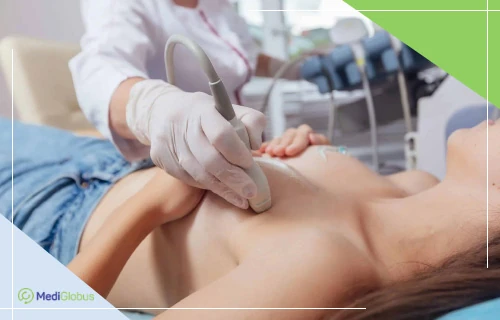
A very fine needle is used to collect the tissue sample, the same size that is used to perform a blood test. The doctor may administer a local anaesthetic before the procedure begins. If the neoplasm is close to the surface, the doctor may immediately aspirate the area; otherwise, ultrasound is used to guide the needle. The doctor makes several punctures at different angles to collect as heterogeneous a tissue sample as possible. Sometimes a fine-needle biopsy is performed to examine fluid-filled cysts; in such cases, it is preferable to collect all of it. After this quick procedure, the doctor applies a sterile dressing – no sutures are required – and the pathological samples are sent immediately to the laboratory for analysis.
Fine-needle biopsy is most commonly used to check large cysts that present with symptoms such as sensitivity in the area of the tumour. It may also be ordered if previous imaging tests have shown vegetations or imaging abnormalities.
Among all the varieties of breast biopsy, fine needle biopsy is the cheapest and least invasive option. According to studies, if a minimum of three tissue samples are taken during the study, the accuracy of the results reaches 91% and the specificity reaches 56%.
However, despite these advantages, many mammologists favour other types of biopsies. When using fine-needle biopsy, there is a higher risk of getting equivocal results, which will require a repeat examination – usually a vacuum or core biopsy. In addition, this diagnostic option is less reliable in distinguishing invasive malignant carcinoma of the breast from an in-situ tumour.
Fine-needle biopsy is considered a relatively easy procedure, and it rarely requires hospitalisation. Possible complications include bruising or haematoma in the injection area, sometimes it can be painful. There is also a risk of infection and pneumothorax if the needle goes too deep into the chest.
 Read:
Read: Core biopsy of the breast
Core biopsy, also known as trepan biopsy, is another common method of diagnosing breast disease.
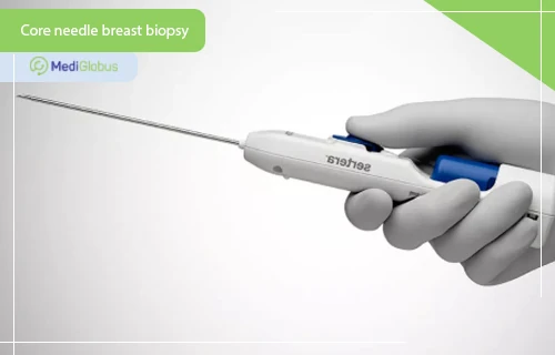
A trepan biopsy uses a special ‘medical gun’ with a hollow needle that collects the tissue. This procedure is carried out under local anaesthesia. Sometimes the doctor may need to make an incision of no more than 0,5 cm to get better access to the tumour. The procedure itself is very quick.
Ultrasound, mammography or MRI may be used to properly guide the needle. The choice of method depends on the specific goals of the doctor – different types of tumours may be better or worse seen depending on which imaging modality is used. Most likely, the patient will be asked to lie on her stomach and her breasts will be compressed during the biopsy. Also, when MRI is used, contrast – an intravenous substance that will help ‘light up’ the tumour on the MRI images – is most often used.
The possibility of performing a biopsy under MRI control depends on the technical equipment of the hospital. Not all medical centres can offer this procedure. Ask the MediGlobus coordinating physicians about the best place to go first.
Compared to fine-needle biopsy, the doctor extracts a larger sample of tumour tissue called a ‘core’, hence the name of the technique. Sometimes the doctor will perform several of these ‘shots’ to collect enough tissue.
Usually, a tiny marker (sometimes called a clip) is implanted in the area from which the tissue samples were taken. It is completely painless and will not be visible from the outside. This is so that during subsequent diagnostics (mammography, MRI, etc.) the doctor can easily and quickly see where the biopsy has already been performed.
In general, the procedure takes 15-30 minutes, if it was carried out without visualisation diagnostics. After that, the doctor puts a sterile dressing on the breast and the patient can go home.
Core biopsy of the breast is considered an accurate and reliable method of diagnosing malignant tumours, which is why mammologists prescribe it when cancer is suspected. It allows them to collect large tissue samples of the neoplasm, thanks to which it becomes possible to carry out additional examinations in the laboratory. Core biopsy also has sufficient sensitivity for difficult-to-detect diagnoses such as invasive lobular carcinoma. Compared to fine-needle biopsy, it results in fewer false-positive and false-negative results.
The disadvantages of core biopsy include worse efficacy in diagnosing women with high breast tissue density. Also, the noise of the gun may be disturbing for some.
Vacuum aspiration breast biopsy
Vacuum-assisted breast biopsy is a newer technique for performing tissue sampling of tumours. It differs from others in that the doctor uses a special imaging device to guide a hollow probe connected to a vacuum device. The procedure is usually performed under ultrasound, MRI or stereotactically.
This method has a very high reliability of results. In many cases, it is superior to fine-needle and core breast biopsy. In particular, patients are less likely to receive false negative results – that is, there is less risk of missing a cancerous process if it is present.
Vacuum aspiration biopsy of the breast is prescribed for:
 The presence of microcalcifications accumulation on previous scans;
Palpable or nonpalpable nodes classified as 3 and 4A according to the American College of Radiologists.
Depending on which imaging technique is used to perform the procedure, the methodology may vary.
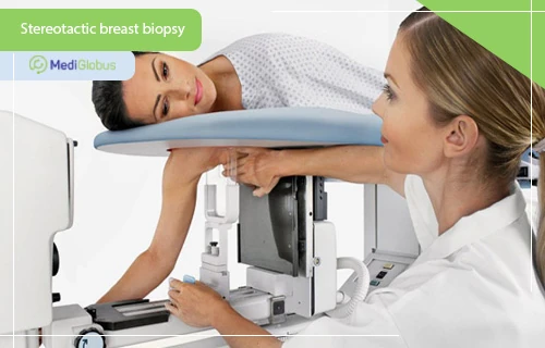
Stereotactic vacuum biopsy
Stereotactic vacuum biopsy is indicated for nodules that appear as microcalcifications on mammography but are not visible on ultrasound. In cases where they are distant from each other, the needle can be withdrawn and reinserted to remove lesions in different areas.
This biopsy technique allows many samples of tumour tissue to be obtained through a single incision. Doctors are able to collect 10 to 20 of these samples per examination, totalling 1,000 to 2,000 mg.
90% of patients who had a breast biopsy said they would prefer stereotactic vacuum-assisted biopsy to open surgery (excisional biopsy).
Ultrasound-guided vacuum-assisted breast biopsy
This version of the procedure uses a special hand-held device that helps the doctor to better visualise the problem area and guide the needle correctly to collect the maximum amount of tumour tissue.
This procedure uses a larger needle. It allows two to three times more tumour tissue to be collected and also makes the biopsy twice as short. Such features of the intervention reduce the likelihood of medical error.
Vacuum breast biopsy has a better ability to obtain fatty breast tissue compared to core biopsy. In addition, vacuum biopsy can be performed on patients with breast implants.
MRI-guided vacuum-assisted breast biopsy
The advantage of using MRI when cancer is suspected is that CT scans can detect tumours even when they are not visible on mammography, ultrasound or physical examination. However, this method of diagnosis will also be the most expensive, which is why doctors do not prescribe it as a routine procedure.
However, due to the fact that MRI technology is based on the use of magnets, which causes the metal in needles to create image artefacts, this diagnostic option is rarely used in clinics. Still, some medical centres have additional equipment that can make the procedure effective – devices for breast fixation, compression, guiding needles and needles made of non-ferrous metals.
Complications of vacuum breast biopsy include bleeding or pain during the procedure; and postoperative pain, bleeding and haematomas. Postoperative compresses and bed rest are effective for their treatment.
Thick-needle (tru-cut) breast biopsy
Today, tru-cut biopsy of the breast is considered to be outdated. It was used in the 80s-90s until it was replaced by fine-needle biopsy. Although its diagnostic capabilities were optimistic, only experienced diagnosticians in specialised clinics were able to achieve optimal results. In addition, the technique had a high false-negative rate in carcinomas and some types of invasive breast cancer.
Excisional (surgical) breast biopsy
Excisional biopsy is a procedure in which the tumour is completely removed during surgery. Like a tri-cut biopsy, it has almost fallen out of use because doctors are usually unable to achieve ‘clean’ resection margins, which is important for a positive cancer treatment outcome. Still, there are a few indications in which surgical biopsy may be considered: atypical lobular hyperplasia, atypical ductal hyperplasia, lobular carcinoma in situ; and in cases where imaging studies disagree significantly with the biopsy, even when repeated.
Punch biopsy of the breast
A punch biopsy is used in specific situations: if cancer is located directly on the skin of the breast. This can be in melanoma, inflammatory breast cancer or Paget’s disease.
A hollow round scalpel called a punch is used to perform the biopsy. On average, the procedure takes about 20 minutes. After the tissue is removed, stitches will be placed in the area. Bruising should go away within 1 to 2 weeks.
Biopsy of a signal lymph node in the breast
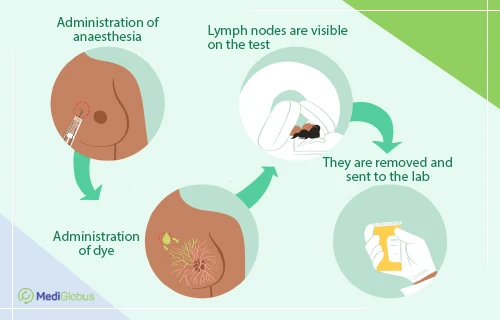
The signal lymph node is the first lymph node to receive drainage directly from the tumour. Its detection and pathological examination can potentially alter the extent and radicality of oncological surgery.
Before the procedure, a staining agent is injected into the patient to allow doctors to correctly identify the right lymph node. The surgeon removes it through a small percutaneous incision.
A signal lymph node biopsy is indicated for patients with early invasive T1-2 breast cancer and clinically negative axillary nodes regardless of breast surgery. Women with ductal carcinoma in situ who have undergone mastectomy are also candidates for it. Sentinel lymph node biopsy may also be considered in patients with ipsilateral recurrent breast cancer after conservative therapy or recurrence after mastectomy.
Frequently asked questions and breast biopsies
Is a breast biopsy harmful?
Performing a breast biopsy is not a health hazard. However, it is an unpleasant procedure, so doctors do not prescribe it without need. In most cases, patients experience pain and discomfort in the area of the injection, which may require short-term administration of mild painkillers. The risk of infection of the biopsy site is very low, and any bleeding is usually minor and goes away on its own.
What diseases can a breast biopsy diagnose?
In Western countries, one in eight women will develop breast cancer at some point in their lives, and about 60% will develop benign breast diseases. Among the latter, fibroadenoma and fibrocystic fibrosis are the most common.
What are the chances that a biopsy will come back positive for cancer?
60-70% of patients who are scheduled for a breast biopsy get results indicating no cancer.
How many tumour tissue samples are taken in a biopsy?
In a standard biopsy, the doctor collects 8-10 samples of tumour tissue. In more specialised techniques, such as vacuum biopsy, the number of samples may be as high as 20.
Can a breast biopsy be performed during pregnancy?
In general, a biopsy is considered a safe procedure that can be performed even in pregnancy.
Where to treat breast cancer?
Based on audits conducted by our doctors, MediGlobus can recommend the following clinics as highly-ranked breast cancer treatment centres. When compiling the ranking, we take into account many factors such as the quality of diagnosis, the technical equipment of the clinic, the availability of all necessary certificates, the professionalism of senior and junior staff, the availability of modern diagnostic and treatment techniques, as well as individual feedback from our patients.
Prof Fatih Agalar: ‘In my practice, we never perform mastectomy (breast removal) in the early stages of cancer. In every situation, we try to preserve the organ as much as possible, and if possible we perform organ-preserving surgery. I recommend my patients who need a mastectomy to undergo breast removal with reconstruction. This operation will cost less and has fewer risks than two separate interventions’.





Summary
A breast biopsy is a diagnostic technique in which the doctor collects a sample of tumour tissue for laboratory examination to look for cancer cells. This procedure is an important step in the diagnosis of malignant and benign breast tumours.
There are different types of breast biopsies. The most common are fine-needle (FNA) and core biopsy. A more modern method is vacuum aspiration breast biopsy. Also among the less common types of biopsy are punch biopsy, excisional biopsy and signal lymph node biopsy, which are performed for specific indications.
Breast biopsy is a safe procedure with a low risk of complications. Pain and bruising will go away within 1-2 weeks.
Provided the diagnosis is carried out in a qualified cancer centre, the probability of a correct diagnosis can reach 99%.
In 60-70% of breast biopsy results for cancer come back negative.
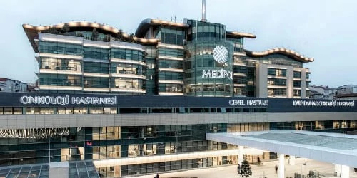
Among the medical centres we recommend for breast cancer patients are Medipol and Liv Vadistanbul (Turkey), Teknon (Spain), Vienna Private Clinic (Austria), Samsung Kangbuk Clinic (South Korea) and Fuda Oncology Centre (China).

Sources:
- 1. Frontiers in Oncology: Look how far we have come: BREAST cancer detection education on the international stage
- 2. American Cancer Society
- 4. StatPearls: Fine Needle Aspiration of Breast Masses
- 5. Gland Surgery: Vacuum-assisted breast biopsy for breast cancer
- 6. Anticancer Research: Stereotactic vacuum-assisted breast biopsy (VABB)–a patients’ survey
- 7. Indian Journal of Surgical Oncology: Why Should We Avoid Excisional Biopsy of Breast Lump? ASOMA Guide for Managing a Discrete Breast Lump
- 8. Irish Cancer Society
- 9. The Cancer Journal: Sentinel Lymph Node Biopsy in Breast Cancer: A Work in Progress