
Basalioma accounts for about 32% of all cancer cases in the world. The disease is also called basal cell carcinoma. It accounts for over 80% of all skin malignancies. Timely diagnosis and proper treatment allow getting rid of this disease completely. Learn more about how to fight against basalioma abroad and how effective it is.
What is basalioma?
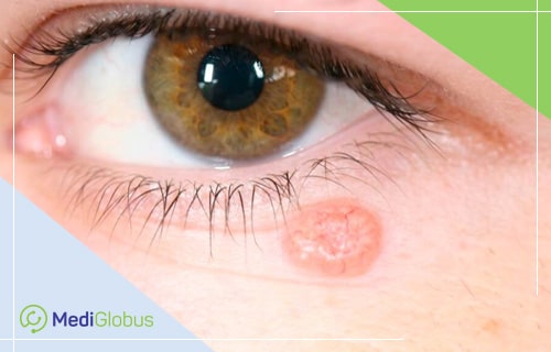
Basalioma is a malignant disease of the basal layer of the epidermis. It is the most common form of skin cancer. A tumour rarely produces metastases. Only in 0,3 cases, it spreads to other organs – bones, lungs and liver.
Basal cell cancer grows slowly. It spreads to neighbouring tissues and destroys them. The neoplasm looks like a shiny pearl-like nodule on the skin or an eczema-like red spot. Some types of basal cell cancer manifest themselves as a thickening on the skin or scar tissue.
The basalioma is caused by DNA damage in the basal cells of the skin. Exposure to ultraviolet radiation from the sun is the main cause of the disease. Ultraviolet is especially harmful in childhood.
Other factors that increase the risk of tumour development include:
![]() prolonged exposure to arsenic;
prolonged exposure to arsenic;
![]() immune system dysfunction;
immune system dysfunction;
![]() radiation therapy;
radiation therapy;
![]() age over 50 years;
age over 50 years;
![]() light skin;
light skin;
![]() chronic skin infections and inflammations.
chronic skin infections and inflammations.
Basal cell cancer occurs in open areas of the body such as the face, ears, neck, lips and back of the hands. Men are more prone to this disease than women.
Types of basalioma
Nodular basal cell carcinoma | Nodular basal cell carcinoma accounts for about 60-80% of all cases. It is most commonly found on the scalp. Clinically, the tumour is represented by elevated pearl-shaped nodes. Over time, ulcers and tissue necrosis appear on such neoplasms. They resemble hemangioma or melanoma, especially if pigmented. |
Cystic basalioma | Appears as one or more cystic nodes of different sizes. Located on the head, neck or back. Usually has a reddish shade. |
Scleroderma form tumor | Tumour cell clusters that have infiltration plaques with a slightly shiny surface and fuzzy borders. Such basalioma occurs on the face or neck. |
Infiltrating basal cell cancer | Typically grows deep inside the skin. The neoplasm has whitish, poorly expressed plaque. The most common localization is in the upper part of the torso or face. After surgical removal it often relapses. |
Micronodular basal cell carcinoma | Such neoplasms are flat or rise slightly above the surface of the skin. They rarely appear and are yellowish-white. The borders are clear and tightened on touch. The most common localization is back skin. |
Superficial basalioma | A tumour ranging in size from a few millimetres to 10 cm or more. This cancer accounts for about 30% of all cases. The neoplasm has a flat-celled plaque with clear borders, a pearl edge and surface erosion. There are no tendencies for invasive growth. Numerous surface basaliomas appear due to arsenic exposure. |
Basalioma diagnostics
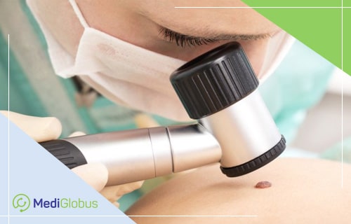
Basalioma is confirmed through a biopsy. The most common method is excision biopsy, performed under local anaesthesia. Tumour tissues are dissected using a scalpel or razor with a curved blade. The obtained samples are examined under a microscope in the laboratory.
Most nodular basal cell tumours of the skin are diagnosed clinically. However, some forms of sociopathology are very difficult to distinguish from hypertrophic scars after acne or benign tumours such as intracutaneous nevus and fibrous papules. In this case, exfoliative cytology methods are used. They have high sensitivity and specificity to confirm the diagnosis.
Basal cell carcinoma stages
Stage 0 | Also called “in situ” carcinoma. Cancer found at this stage is only present in the upper layer of the skin and does not spread deeper into the dermis. |
Stage 1 | Tumour size 2 cm or less, no metastases. Lymph nodes are not affected. |
Stage 2 | A tumour larger than 2 cm. It has not spread to nearby organs and lymph nodes. |
Stage 3 | Any size of a tumour. A single lymph node measuring 3 cm or less is involved in the malignant process. No metastases. |
Stage 4 | Tumour of any size. Two or more lymph nodes are affected. Multiple remote metastases present. |
Methods of basal cellular skin cancer treatment
Surgical treatment of basalioma
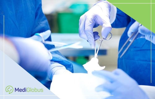
Surgery is the main treatment option. The neoplasm is dissected together with 3-10 mm of healthy tissue.
According to the National Center for Biotechnology Information, after surgical removal of basalioma up to 1.5 cm in diameter, the risk of relapse within 5 years is about 10%. For tumours larger than 3 cm, this figure exceeds 23%.
It is believed that relapses arise from residual cancer cells that are not always detected by conventional histology. Primary tumours of the ear auricle, eyelid, scalp and nose recur in 12-25% of patients.
Mohs surgery
It’s a microsurgical intervention that involves layer-by-layer removal of the tumour. Each section of the neoplasm undergoes a histological check. The main purpose of the surgery is to keep the tissue as healthy as possible and eliminate the risk of relapse. The intervention is used for primary basaliomas with complex localizations (nose, lips, eyelids, sexual organs) or with a high risk of recurrence. Also, tumours with fuzzy edges and a diameter greater than 2 cm are treated with microscopic surgery.
X-ray therapy
The doctor uses X-rays to destroy the malignant focus. The procedure is performed without anaesthesia. The patient may need to undergo several repetitions to destroy cancer cells over a couple of weeks or daily procedures for 5-7 days. The risk of recurrence is only 6.3%. Radiotherapy is indicated for tumours up to 5 cm in size on the nose and nasolabial folds in elderly patients.
Electrocoagulation and scraping of basalioma
The doctor scrapes the tumour using a curette. The surgeon uses a special chemical to destroy the remaining cancer cells, stop the bleeding and seal the wound. It’s possible to repeat the procedure during one session.
The operation leaves a round whitish scar, reminiscent of a cigarette burn.
The method applies to surface tumours from 2 to 5 cm in diameter. After treatment, the relapse rate varies from 1 to 15% for small neoplasms and increases to up to 50% for basalic tumours over 3 cm in diameter.
Cryotherapy for basalioma
This method is used for tumours up to 2 cm in size with clear boundaries. Cryotherapy is used on patients who have contraindications for surgical removal. The frequency of relapse after the treatment of small (up to 8 mm) primary tumours is about 8%. However, these patients need to be closely monitored. Specific contraindications are localizations in the nasolabial fold, on the nose, on the ear tragus, at the edge of the eyelid.
During the procedure, the doctor uses a cotton swab applicator or atomiser to apply liquid nitrogen. This makes it possible to freeze and destroy cancer cells. The treated areas are covered with blisters or scabs, under which healthy skin appears.
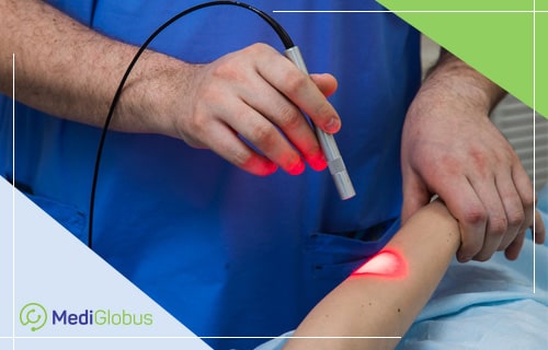
Laser treatment of basal cell skin cancer
Suitable for treating surface tumours. According to the National Center for Biotechnology Information, the relapse rate after laser therapy ranges from 3.7 to 15.5%.
Laser therapy uses laser beams for the destruction of cancer cells. This makes it possible to destroy the tumour without damaging the healthy tissue around it. The healing of the wound goes as fast as possible.
Photodynamic therapy for basalioma
During the procedure, a special drug is used, which makes the tumour sensitive to light. It is injected into the basalioma or applied to its surface. Then the cancer cells are destroyed with a photodynamic apparatus.
Patients should avoid sunlight for at least 48 hours after the procedure. As UV exposure increases the activation of the drug and causes serious sunburn.
This method is used to treat single or multiple surface basalis. The risk of recurrence after photodynamic therapy is about 15%.
Medication therapy for basal cell cancer
Medication therapy is used when surgical treatment is impossible. The Food and Drug Administration (FDA) has approved two oral medications to treat progressive basalioma in adults. With these drugs, it is possible to fight tumours that have penetrated deep into the skin, spread to other parts of the body and are not amenable to other oncological methods. Both drugs block the development of the malignant process and destroy cancer cells.
Basalioma treatment prognosis
The prognosis for cancer patients with basal cellular skin cancer is favourable. For the localized tumours of the initial stages, the 5-year survival rate of patients is 95-99%. For stage 4 of the disease, this index does not exceed 10%. It should be noted that most cases are diagnosed at the initial stages of development.
Resume
![]() Basalioma is common skin cancer. The tumour is successfully diagnosed abroad. Early detection of pathology and adequate treatment can completely get rid of the disease.
Basalioma is common skin cancer. The tumour is successfully diagnosed abroad. Early detection of pathology and adequate treatment can completely get rid of the disease.
![]() Despite its slow growth and numerous therapeutic methods, basalioma should not be underestimated. The main treatment method is surgery. The risk of relapse after such treatment is extremely low. The number is less than 10% in the initial stages and about 23% in the late stages.
Despite its slow growth and numerous therapeutic methods, basalioma should not be underestimated. The main treatment method is surgery. The risk of relapse after such treatment is extremely low. The number is less than 10% in the initial stages and about 23% in the late stages.
![]() The prospects for people with basalioma are good. Almost every case of sociopathology can be completely cured, especially if it is detected at an early stage.
The prospects for people with basalioma are good. Almost every case of sociopathology can be completely cured, especially if it is detected at an early stage.
To go for basalioma treatment abroad, leave your application on the website. Doctors-coordinators of the international MediGlobus platform will help you choose a clinic and organize your medical trip.