
Leiomyosarcoma is one of the rarest types of cancer. It occurs in 0.7% of cancer patients. Leiomyosarcoma is aggressive and difficult to treat, so it requires a reliable diagnosis and rapid treatment.
You can find out about approaches to leiomyosarcoma treatment abroad in this article.
What is leiomyosarcoma?

Leiomyosarcoma is a type of soft tissue cancer. It grows from soft muscles, most commonly in abdominal organs. In half of the cases, leiomyosarcoma is formed inside the retroperitoneal space. A different type of disease is leiomyosarcoma of the uterus, which has a similar origin, but its nuances in diagnostics and treatment.
A tumour can grow up to 10 cm in diameter and cause significant discomfort and pain. Large masses usually cannot be completely surgically removed.
Leiomyosarcoma occurs in people aged 50-60 years, in women twice as often as in men. However, it also can occur in children. People with reduced immunity due to organ transplants or diseases such as AIDS or the Epstein-Barr virus are at increased risk of developing this cancer.
Leiomyosarcoma is divided into four subtypes, depending on the main affected area:
Soft tissue leiomyosarcoma
It’s the most common variety. Most often found in the abdominal area. In most cases, it grows from the muscle tissue of small vessels. Somatic tumours can reach a large size, which is associated with poor prognosis.
Leiomyosarcoma of the skin
It affects the dermis – the middle layer of skin located between subcutaneous fatty tissue and the surface layer of skin. Unlike other subtypes, it is more common in men. Typically, tumours are small, up to 1-2 cm in diameter and have a good prognosis. If the formation grows deeper, it gives metastases in 30-40%, usually in the lungs.
Uterine leiomyosarcoma
Most often found in women 40-60 years old. Makes up 1% of all uterine cancer cases. An aggressive type of cancer, which progresses rapidly. Often the diagnosis is established after the surgery, based on histological analysis of tissue.
Leiomyosarcoma of the vessels
It’s very rare. There are only a few hundred known cases of vascular leiomyosarcomas in the world. Symptoms depend on the location of the tumour. In 54% of cases, liver and lung metastases occur.
Depending on the subtype, the prognosis and treatment strategy differ.
Symptoms of leiomyosarcoma
Leiomyosarcoma has no symptoms to distinguish it from other types of soft tissue sarcoma. As a result, it is often mistaken for other malignancies, most commonly leiomyoma and fibroids. To confirm the diagnosis, histological tissue analysis is always performed.
Leiomyosarcoma of the soft tissue of the abdomen may manifest itself in the following symptoms:
![]() pain;
pain;
![]() swelling;
swelling;
![]() weight loss;
weight loss;
![]() nausea and vomiting.
nausea and vomiting.
Uterine leiomyosarcoma is usually accompanied:
![]() abnormal vaginal bleeding;
abnormal vaginal bleeding;
![]() abdominal pain;
abdominal pain;
![]() weight loss;
weight loss;
![]() weakness.
weakness.
Leiomyosarcoma, located on the vessels, often takes the form of large, painless tumours. In rare cases when large vessels are affected, they may be compressed. This will manifest in symptoms such as swelling and numbness.
Stages of uterine leiomyosarcoma
Stage 1 | The tumour is within the uterine endometrium. |
Stage 2 | The tumour is spreading to the cervix. |
Stage 3 | Extension of the cancer process to the peritoneum, vagina and/or lymph nodes. |
Stage 4 | Spreading of the tumour into the bladder and/or intestinal tissue, formation of remote metastases. |
Stages of soft tissue leiomyosarcoma
Stage 1 | Surface tumour up to 5 cm, low grade. |
Stage 2 | The tumour is more than 5 cm deep, low grade, or less than 5 cm high grade. |
Stage 3 | The tumour is larger than 5 cm, deeply located, high grade. |
Stage 4 | Presence of metastases at any characteristics of the tumour. |
How is leiomyosarcoma diagnosed?
In leiomyosarcoma, as in any other form of aggressive cancer, rapid diagnosis is important. The doctor determines the stage of the disease, as well as the location of the tumour and its borders, which is important in planning the operation. Leiomyosarcoma cannot be diagnosed using imaging techniques alone (MRI, CT, etc.), cell analysis is essential. Often, the diagnosis is confirmed already after the tumour removal operation.
MRI. Magnetic resonance imaging is always done when sarcoma is suspected. With its help, it is possible to estimate the size and location of a tumour and its borders. MRI can also detect if cancer has begun to spread into neighbouring tissues (nerves, bones) or compress vessels.
CT. Often, together with an MRI, doctors do a CT scan. It is especially useful for tumours of the retroperitoneal space and metastases in the abdominal or chest organs, especially the lungs.
Angiography is a method of blood vessel research that uses X-rays. It is effective for the diagnosis of a large vessel tumour.
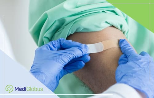
Biopsy. To confirm that the patient has leiomyosarcoma and not another type of cancer, doctors need to examine the tumour cells in the lab. This is done by biopsy, a procedure to extract a sample of tissue from the body. In leiomyosarcoma, a needle biopsy is most often performed using computerized tomography. The patient is placed inside the CT machine and the doctor can see on the monitor screen where exactly he or she sends the needle. This method is more accurate than a conventional biopsy and takes less time.
Prognosis for leiomyosarcoma
It is difficult to make a prognosis for leiomyosarcoma as it is a rare oncological disease and doctors do not have enough data for accurate analysis.
The prognosis in leiomyosarcoma depends on the location of the tumour, the stage of the disease, the degree of differentiation and the presence of metastases.
Leiomyosarcoma is an aggressive type of cancer. 90% of patients are at stage 2 or 3 at the time of initial diagnosis.
Approximate survival rate at leiomyosarcoma uteri, depending on stage:
![]() Stage 1 – 44-64%;
Stage 1 – 44-64%;
![]() Stage 2-3 – 19-53%;
Stage 2-3 – 19-53%;
![]() Stage 4 – 8-39%.
Stage 4 – 8-39%.
The total five-year survival rate for soft tissue sarcoma is 64%.

Methods of treatment of leiomyosarcoma
Given the complexity and aggressiveness of the disease, the most effective treatment for leiomyosarcoma is carried out in centres that specialize in sarcoma. Such institutions usually have modern equipment for an accurate diagnosis. They also employ specialists from different fields – oncology, radiology, surgery, etc. – who have experience in the treatment of this type of tumour.

Leiomyosarcoma surgery
The main method of treatment. The surgeon plans a procedure based on diagnostic tests. Together with the tumour, the healthy tissue edge is removed to prevent a relapse. Depending on the stage, the surgeon can also remove nearby lymph nodes. The extent of the intervention for uterine leiomyosarcoma depends on the stage, but the minimum standard is the complete removal of the uterus and the cervix. For premenopausal patients, the doctor will discuss the possibility of preserving the ovaries. This issue has its advantages and risks, the decision is made on a case-by-case basis.
Radiotherapy
Since tumours are often near vital organs, the complete removal of malignant cells may not be possible. Therefore, in most cases, doctors use radiotherapy together with conventional surgery to reduce the tumour or destroy the remaining cancer cells. Radiotherapy is also often used to stop the spread of metastases. In uterine leiomyosarcoma, radiotherapy is used rarely.
Brachytherapy
In this procedure, the radioactive source is placed inside the body, next to the tumour. This treatment method is most often used for stage 3 uterine leiomyosarcoma, in combination with chemotherapy and radiotherapy.
Chemotherapy
In soft tissue leiomyosarcoma, chemotherapy is usually used to treat metastases. It is also effective in reducing large tumours (over 8 cm) before surgery. In stage 3-4 leiomyosarcoma of the uterus, chemotherapy can be effective in combination with radiotherapy.
After treatment, the patient should be regularly monitored by an oncologist to prevent relapse. It is recommended to have an examination every 3 months during the first two years and then every 6-12 months. Also, a CT/MRI scan is conducted every 3-6 months for the first two years and every year thereafter.
Clinics that specialize in sarcoma treatment
![]() Koç University Hospital (Turkey);
Koç University Hospital (Turkey);
![]() University Clinic of Navarra (Spain);
University Clinic of Navarra (Spain);
![]() Quironsalud Hospitals Group (Spain);
Quironsalud Hospitals Group (Spain);
![]() Ichilov Medical Center (Israel);
Ichilov Medical Center (Israel);
![]() Ludwig-Maximilian Medical Center (Germany);
Ludwig-Maximilian Medical Center (Germany);
![]() Helios Clinic (Germany);
Helios Clinic (Germany);
![]() Soon Chun Hyang Hospital (South Korea);
Soon Chun Hyang Hospital (South Korea);
![]() Anam Clinic (South Korea).
Anam Clinic (South Korea).
Resume
![]() Leiomyosarcoma is an aggressive soft tissue cancer. This type of cancer is prone to rapid progression and the formation of metastases..
Leiomyosarcoma is an aggressive soft tissue cancer. This type of cancer is prone to rapid progression and the formation of metastases..
![]() The average five-year survival rate with leiomyosarcoma is 64%.
The average five-year survival rate with leiomyosarcoma is 64%.
![]() Treatment standards for leiomyosarcoma depend on the stage, size, location and grade of the tumour. Surgical removal is often accompanied by radiotherapy, chemotherapy and brachytherapy.
Treatment standards for leiomyosarcoma depend on the stage, size, location and grade of the tumour. Surgical removal is often accompanied by radiotherapy, chemotherapy and brachytherapy.
![]()
The best results in the treatment of leiomyosarcoma are achieved by specialized oncology centres. Such clinics are present in Germany, Spain, Israel, Turkey and South Korea.
Treatment of leiomyosarcoma should be started without delay. To sign up for a consultation with a foreign specialist, leave your application on the MediGlobus website by clicking on the “Get a free consultation” button. Our coordinating doctors will call you back and help with the organization of treatment.
Получить бесплатную консультацию
