
Visualization diagnostics involves obtaining data on the functioning of internal organs and structures in a convenient form for visual perception. In contrast to invasive examinations, such methods do not involve surgical intervention, and the integrity of the soft and hard tissues of the body is not compromised. In recent years, they have become more informative, accurate and safe for the patient. Learn which diagnostic procedures are the most popular, how they differ, what the pros and cons are, and where they have found their application in this article.
Listen to the article:
The most popular diagnostic methods
Globally, about 5 billion medical imaging procedures were performed until 2010. In the last decade, this number has multiplied.
Modern imaging diagnostics help to make correct diagnoses quickly and without any harm to the body. They have found wide applications in different fields of medicine. Imaging tests are often used to detect cardiovascular diseases, cancer, neurological disorders, abdominal diseases, musculoskeletal abnormalities and other serious diseases.
The most informative and best diagnostic procedures are:
![]() magnetic resonance imaging (MRI),
magnetic resonance imaging (MRI),
![]() positron emission tomography (PET),
positron emission tomography (PET),
![]() computerized tomography (CT),
computerized tomography (CT),
![]() optical tomography of the spine,
optical tomography of the spine,
![]() ultrasonography,
ultrasonography,
![]() single photon emission computed tomography.
single photon emission computed tomography.
To make an appointment with a diagnostic centre abroad, click on the button below and fill out the feedback form. We will help you with organizing a check-up trip and choosing a clinic.
MRI scan

Magnetic resonance imaging (MRI) is one of the most sought-after methods of 3D body imaging. It allows you to use nuclear magnetic resonance to obtain three-dimensional detailed images of internal organs and body structures.
The procedure is actively used in neurology and neurosurgery, oncology, vascular surgery, and other branches of medicine. This examination provides information about the functionality of the brain and spinal cord, as well as soft tissues: muscles, cartilage, and ligaments.
MRI is most commonly prescribed to diagnose abnormalities of the brain, blood vessels, spine, abdominal organs, and thorax. It may be performed to detect diseases of individual organs, such as the intestines, heart, lungs, kidneys, liver, etc.
Pros of MRI:
![]() speed of diagnosis (15 to 60 minutes);
speed of diagnosis (15 to 60 minutes);
![]() helps detect pathology at an early stage;
helps detect pathology at an early stage;
![]() high quality 3D images;
high quality 3D images;
![]() available for diagnosing any organ;
available for diagnosing any organ;
![]() there are no risks of ionizing radiation;
there are no risks of ionizing radiation;
![]() when using an MRI contrast agent, the probability of allergic reaction to it is lower than to CT or X-ray with iodine-based contrast.
when using an MRI contrast agent, the probability of allergic reaction to it is lower than to CT or X-ray with iodine-based contrast.
Cons of MRI:
![]() noise,
noise,
![]() may cause fear of confined space during scanning,
may cause fear of confined space during scanning,
![]() examination of the skeleton is ineffective (CT or X-ray is better for this purpose),
examination of the skeleton is ineffective (CT or X-ray is better for this purpose),
![]() you cannot move during the procedure.
you cannot move during the procedure.
Modern MRI machines
Abroad, MRI scans are performed on the latest generation of CT scanners. These machines differ for the better from those that are mostly used in the former post-Soviet countries.
These units make it possible to speed up the scanning process without reducing the quality of the resulting images. They are equipped with special StarVIBE image clarity correction systems. This is necessary to avoid artefacts appearing on MRI scans due to involuntary human movements (e.g. swallowing or breathing).
This equipment also has Innovision’s infotainment system. It includes comfortable memory foam cushions that reduce scanning noise and provide clear audio cues to the patient.
Innovision immerses patients in a world of music and video. It informs them about the remaining scanning time. Another big benefit: The display makes the scanner opening appear large, which prevents the patient from feeling claustrophobic.
Modern magnetic resonance imaging machines are designed to diagnose people with high body weights and deformed skeletal structures. Thanks to the soft, ultralight coils and Quiet Suite positioning aids, the patient can relax and take the most comfortable position possible.
Some MRI scanners have an open tunnel design, which is ideal for children and people with a fear of confined spaces. Also, the opening of newer models of MRI scanners is 70-80 cm in diameter. This is about 20% larger than in the old models.
PET scan
Positron emission tomography (PET) is a nuclear diagnostic procedure that allows an accurate assessment of the biochemical and metabolic functions of the human body. This examination is often combined with CT or MRI, in which case it is referred to as PET-CT or PET-MRI.
The technique is based on the detection of radioactive signals emitted by tissue after a small amount of radioactive indicator is injected intravenously into the patient’s bloodstream. It is most commonly used to detect diseases of the brain, heart or cancer. PET scans can also be used to check the entire body.
Advantages of PET
![]() better sensitivity than other imaging tests (CT, MRI),
better sensitivity than other imaging tests (CT, MRI),
![]() diagnostic accuracy,
diagnostic accuracy,
![]() takes about 30 minutes,
takes about 30 minutes,
![]() painlessness,
painlessness,
![]() wide area of application.
wide area of application.
Disadvantages of PET
![]() radiation exposure,
radiation exposure,
![]() may give false results if the chemical balance in the body is not normal,
may give false results if the chemical balance in the body is not normal,
![]() cost.
cost.
New-generation positron emission tomography devices are highly sensitive biomedical imaging devices. They are equipped with SiPM PET systems, which provide high-quality three-dimensional images of pathological tissues, particularly tumours at the earliest “in situ” stage. These units also have programs for correcting scans with involuntary movements of the patient. This helps to obtain clear images without artefacts and blurring.
Computed tomography scan

Computed tomography (CT) is a diagnostic procedure that uses X-ray equipment and computers to create cross-sectional images of the body. It clearly shows the condition of internal organs, blood vessels, bones, and soft tissue.
The examination is used to detect abnormalities of the cardiovascular system, disorders of the musculoskeletal system, lung diseases, oncology, problems of the urogenital system, dental diseases, etc. It can also be used to visualize the process during puncture or some surgical interventions.
CT diagnostics abroad are performed with the latest models of CT scanners. Unlike their predecessors, which made 32, 64, and 128 slices per resolution, the new generations of these machines can make 512 and 640 slices. They produce high-quality images without any distortions.
CT strengths:
![]() Speed of procedure (5-15 minutes),
Speed of procedure (5-15 minutes),
![]() Image quality,
Image quality,
![]() Fast results (30-50 minutes),
Fast results (30-50 minutes),
![]() Absence of long preparation for the procedure,
Absence of long preparation for the procedure,
![]() Painlessness.
Painlessness.
Weaknesses of CT:
![]() Ionizing radiation,
Ionizing radiation,
![]() Risks of allergic reaction to the contrast agent,
Risks of allergic reaction to the contrast agent,
![]() Low natural contrast of soft tissues.
Low natural contrast of soft tissues.
CT and alternative diagnostic methods
CT and MRI have a similar principles of operation. These two methods create three-dimensional images from multiple scans taken from different angles. But CT uses X-rays, and MRI uses electromagnetic waves. The first procedure is more informative for diagnosing bones, blood vessels, lungs, teeth, and heart, while the second is more informative for soft tissues and tumours.
A CT scan may be recommended instead of an MRI if the patient has a metal implant, pacemaker or another device that is contraindicated for an MRI. Unlike a simple x-ray, a CT scan offers a much higher level of detail in the areas being scanned, creating clear, computerized 360-degree images of body structures.
Ultrasound diagnostics

Ultrasound is a non-invasive way of diagnosing the body. It is one of the safest and simplest methods of obtaining layer-by-layer images of tissues and organs. It is used in various areas of medicine for patients of all ages.
Ultrasound is an ideal way to assess the condition of the fetus, examine internal organs, perform a needle biopsy, or detect any vascular abnormalities. The examination is also called sonography. During it, a special sensor sends sound waves that pass through the patient’s body and cause an echo. This data is displayed on a computer screen.
Modern ultrasound scanners have a high resolution, which allows you to get detailed information about the areas being examined. They are equipped with 3D and 4D technologies, which provide three-dimensional images in real-time and therefore clearer images.
Advantages of ultrasound
![]() Painless and safe (can be performed on pregnant women and newborns),
Painless and safe (can be performed on pregnant women and newborns),
![]() Low cost,
Low cost,
![]() Affordability,
Affordability,
![]() Mobility (there are portable ultrasound machines that can be brought to the patient’s room to conduct the examination),
Mobility (there are portable ultrasound machines that can be brought to the patient’s room to conduct the examination),
![]() Clear images of soft tissues,
Clear images of soft tissues,
![]() No radiation exposure.
No radiation exposure.
Disadvantages of ultrasound
![]() A more superficial examination than, for example, an MRI or CT scan,
A more superficial examination than, for example, an MRI or CT scan,
![]() Poor penetration of ultrasound waves through bone or air,
Poor penetration of ultrasound waves through bone or air,
![]() The quality of the results depends on the equipment and the skills of the doctor,
The quality of the results depends on the equipment and the skills of the doctor,
![]() May be insufficiently informative in people with significant obesity.
May be insufficiently informative in people with significant obesity.
It is expected that modern ultrasound machines will soon be able to support artificial intelligence. This system will allow doctors to interpret images quickly and accurately, as well as to identify patterns that a doctor might miss in a conventional scan.
Optical topography of the spine
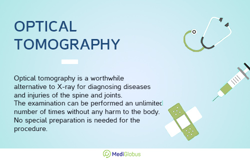
Computerized optical tomography is a worthy alternative to X-ray methods for diagnosing diseases and injuries of the spine and joints. It can easily detect scoliosis, kyphosis, lordosis, pelvic misalignment, and other pathologies.
The DIERS device is used to perform optical tomography. It consists of a special treadmill, optical systems and a computer.
During the session, the patient stands or moves on a barometric platform while the optical camera feeds horizontal “light” strips into the back area. Thanks to the stereo effect, this creates 3D and 4D images of the spinal column. Therefore, the doctor can view the examined area from all angles in a three-dimensional image. After the procedure, the computer program processes the information and gives the result.
Strengths of optical tomography
![]() high accuracy,
high accuracy,
![]() no radiation exposure,
no radiation exposure,
![]() painlessness,
painlessness,
![]() informativeness,
informativeness,
![]() the rapidity of the study (5-10 minutes),
the rapidity of the study (5-10 minutes),
![]() availability.
availability.
Weaknesses of optical tomography
![]() Limitation of the patient’s weight and height (not suitable for patients who are shorter than 90 cm or taller than 210 cm, or for people weighing more than 200 kg),
Limitation of the patient’s weight and height (not suitable for patients who are shorter than 90 cm or taller than 210 cm, or for people weighing more than 200 kg),
![]() May not be informative in patients with large back tattoos or skin that is too dark.
May not be informative in patients with large back tattoos or skin that is too dark.
Optical tomography not only allows you to detect diseases but also to monitor the effectiveness of treatment. It can be performed an unlimited number of times without any harm to the body. No special preparation is needed for this examination.
Single Photon Emission Computed Tomography (SPECT)

Single-photon emission computed tomography is one of the most popular radionuclide diagnostic techniques. It is similar to PET. The procedure uses a radioactive substance that is injected intravenously and a special gamma camera to create 3D images.
SPECT is mainly prescribed for the diagnosis and monitoring of brain, heart, liver, bone, and breast pathologies. It is recommended for various neuropsychiatric disorders resulting from chemical imbalances in the brain (e.g., Alzheimer’s disease, depression, schizophrenia, bipolar disorder, Parkinson’s disease).
Unlike other imaging tests, such as X-rays, which show what the structures inside the body look like, SPECT creates images of exactly how organs function.
For example, this method can see if blood flow to the heart is impaired, which areas of the person’s brain are more or less active, and how much and which bone tissues are affected by the malignant process.
Pros of SPECT
![]() high sensitivity,
high sensitivity,
![]() ability to scan the entire body as well as individual body parts and organs,
ability to scan the entire body as well as individual body parts and organs,
![]() displays several biological processes,
displays several biological processes,
![]() can be used in the presence of electronic devices implanted in the heart,
can be used in the presence of electronic devices implanted in the heart,
![]() cost.
cost.
Cons of SPECT
![]() Long scanning time,
Long scanning time,
![]() low-resolution images,
low-resolution images,
![]() possibility of artefacts in tomograms,
possibility of artefacts in tomograms,
![]() does not provide a quantitative assessment of blood flow,
does not provide a quantitative assessment of blood flow,
![]() has some radiation risk, although very low.
has some radiation risk, although very low.
SPECT costs less than PET but produces lower-resolution images. However, this lower resolution can be used perfectly for a variety of medical purposes. And the fact that the procedure costs less than positron emission tomography makes it more affordable.
Today the field of nuclear medicine is increasingly using SPECT/CT. This is a hybrid version of SPECT and CT. It provides accurate information about bone and soft tissue conditions for inflammation, tumours, infection, and trauma of the musculoskeletal system. SPECT/CT also helps in therapy planning.
Summary
![]() There are many imaging diagnostic methods available. They help to detect abnormalities of internal organs, the brain, circulatory or nervous system, etc. More than 5 billion such procedures have been performed worldwide.
There are many imaging diagnostic methods available. They help to detect abnormalities of internal organs, the brain, circulatory or nervous system, etc. More than 5 billion such procedures have been performed worldwide.
![]() Among the most popular medical imaging techniques are positron emission tomography, ultrasonography, optical tomography, single-photon emission computed tomography, and magnetic resonance and computed tomography.
Among the most popular medical imaging techniques are positron emission tomography, ultrasonography, optical tomography, single-photon emission computed tomography, and magnetic resonance and computed tomography.
![]() Leading clinics abroad use the latest modifications of CT, MRI, ultrasound, PET and other examinations. Such equipment allows for the most comfortable, safe and high-quality examinations.
Leading clinics abroad use the latest modifications of CT, MRI, ultrasound, PET and other examinations. Such equipment allows for the most comfortable, safe and high-quality examinations.
To travel to one of the best medical centres abroad for a diagnosis, contact the MediGlobus coordinating physicians. We will assist the patient every step of the way in organizing the trip.

Sources:
- National Library of Medicine
- Montgomery Radiology Associates
- American Society of Clinical Oncology (ASCO)
- Mayo Clinic
- ScienceDirect
- Springer Nature Switzerland AG
- National Library of Medicine

